Research Article
Marvin Rubenstein, PhD 1, 2, 3, 4*, Courtney M.P. Hollowell, MD 4, Patrick Guinan, MD 1, 3, 4, 5
1Division of Cellular Biology, Hektoen Institute for Medical Research, Chicago, IL, 60612, USA
2Department of Biochemistry, Rush University Medical Center, Chicago, IL, 60612, USA
3Department of Urology, Rush University Medical Center
4Division of Urology, Stroger Hospital of Cook County, Chicago, IL, 60612, US
5Department of Urology, University of Illinois at Chicago, Chicago, IL, 60612, USA
Corresponding author
Dr. Marvin Rubenstein, Chairman, Division of Cellular Biology, Hektoen Institute for Medical Research, 2240 West Ogden Avenue, Chicago, IL, 60612, USA, Tel: +1 312 864 4621; Fax: +1 312 768 6010; E-mail: DrMarv@Prodigy.net
Received Date: 23 Jan 2014
Accepted Date: 27 Apr 2014
Published Date: 05 May 2014
Citation
Rubenstein M, Hollowell CM, Guinan P (2014) In Lncap Cells Inhibition of Bcl-2 by Antisense Oligonucleotides Results in Compensatory Changes Involving Apoptosis, Transcription and Immunoregulation. Enliven: J Genet Mol Cell Biol 1(2): 001.
Copyright
@ 2014 Dr. Marvin Rubenstein. This is an Open Access article published and distributed under the terms of the Creative Commons Attribution License, that permits unrestricted use, distribution and reproduction in any medium, provided the original author and source are credited.
Abstract
Antisense oligonucleotides (oligos) have been evaluated for treating prostate cancer in both in vivo and in vitro models. Although most oligos contain
a single mRNA binding site, our laboratory evaluates bi-specifics directed towards two proteins. This study evaluates the growth inhibition of in vitro
propagating LNCaP cells employing mono- and bi-specific oligos directed against BCL-2 [the second binding site was directed against the epidermal
growth factor receptor (EGFR)]; and employing RT-PCR, the expression of five apoptosis regulatory proteins (BCL-2, bax, caspase-3, clusterin, AKT-
1), a tumor associated transcription factor (MED-12) and an immune blockade associated regulatory marker (PD-L1) were evaluated.
LNCaP prostate tumor cells were incubated in the presence of oligos specifically directed against BCL-2 (entering the cells through a form of
nanodelivery) and compared to lipofectin containing controls. Significant, but comparable, growth inhibition was produced by both mono- and bispecific
forms. Employing RT-PCR to determine BCL-2 expression, we found that the greatest amount of mRNA suppression approached 100% for
each type of oligo: mono-specific MR4 (directed only against BCL-2), 100%; and bispecifics MR24 and MR42, 86% and 100% respectively. Based upon
both inhibition of in vitro growth and BCL-2 expression, bi-specific antisense oligos directed against EGFR and BCL-2 mRNAs are at least as effective
as a mono-specific directed solely towards BCL-2.
The objective of these experiments was to determine a compensatory response by cells to (again) evade apoptosis in the presence of BCL-2 suppression. Levels of mRNA encoding non-targeted bax, caspase-3, clusterin and AKT-1 were initially evaluated, while additional experiments sought to identify additional mechanisms of tumor adaptability and resistance. Suppression of the apoptosis inhibitor (BCL-2) in LNCaP cells did not alter either bax or clusterin expression. However, non-targeted caspase-3 (an apoptosis promoter) was suppressed and non-targeted AKT-1 (an apoptosis inhibitor) was enhanced. This suggests that tumor variants can resist apoptosis through the altered expression of non-targeted regulators of apoptosis. Additional
experiments identified other mechanisms of compensation involving transcription and immune regulation suggesting further studies are needed.
Keywords:
Antisense oligonucleotides; Prostate cancer; BCL-2; Bax; Caspase-3; Clusterin; AKT-1; MED-12; PD-L1; Therapy
Introduction
Antisense oligonucleotides (oligos) have been evaluated in both in vivo and in vitro prostate cancer models primarily through suppression of protein growth factors, androgens (oligos directed against 5-alpha-reductase), receptors which bind these stimulating factors, regulators of apoptosis and oncogenes. Those which have advanced to clinical trials focus on the inhibitors of apoptosis (BCL-2 [directly an inhibitor] and clusterin [an inhibitor of activated bax, which is a promoter]). Oligos provide a specific, relatively non-toxic method for translational arrest, and almost all are directed against single gene products or those which shared sequence homology. Since most tumors are characterized by the enhanced expression of many proteins, growth is unlikely to be inhibited by knockdown of single gene products. Instead, multiple genes must be down regulated. Therefore, our laboratory evaluates bi-specific oligos which target two distinct proteins (able to regulate different pathways and which do not share sequence homology). These bi-specifics differ from those targeting genes which share sequence homology (BCL-2 and BCL-xL) [1] or the OGX-225 oligo which targets (three) structurally related insulin like growth factor binding proteins [2]. We have demonstrated that the addition of a second binding site does not affect the activity of the first and that dual binding sites can simultaneously be directed against genes involved in either a single growth promoting autocrine loop [3] or towards those of even different regulatory pathways [4-7]. While most assessments of activity quantitate the inhibition of in vitro growth, more specific methods use the polymerase chain reaction (PCR) to measure specific protein encoding mRNA.
Previously we have shown that in LNCaP cells a single mono- and two bispecific oligos directed towards BCL-2 increase the expression of the androgen receptor (AR) [8] and its co-activating transcription factors p300 and interleukin-6 (IL-6) [9]. Since this pattern of gene expression is often associated with later stage tumors, we postulated that suppressive therapy against BCL-2 could lead to increased androgen sensitivity and promote tumor aggressiveness. It is likely that additional compensatory mechanisms exist, and since regulation of apoptosis is clinically receptive to oligo treatment, we evaluated several non-targeted proteins of this process to identify other proteins associated with compensation and apoptosis evasion. We used the same mono- and bi-specific oligos as in the androgen sensitivity studies and RT-PCR to evaluate BCL-2, bax, caspase-3, clusterin and AKT-1. These proteins were chosen because they regulate apoptosis in opposing manners; BCL-2, clusterin and AKT-1 are inhibitory, while activated bax and caspase-3 stimulate the process. MED-12 is a protein which acts as a mediator for RNA polymerase and mutated forms are associated with prostate cancer [10]. PD-L1 is the ligand for the PD (programmed death) receptor expressed on cytotoxic T cells and is currently being evaluated in immune blockade therapy for melanoma and other solid tumors [11].
If gene therapy is to be effective when directed against BCL-2, compensatory changes which compromise the effectiveness of the desired suppression must be identified. Among a mass of heterogeneous tumor cells, those which evade apoptosis are most likely to be selected and altered patterns of gene expression could include increased inhibition by clusterin or AKT-1 or decreased promotion by bax and caspase-3. In addition alterations in transcription or immunoregulation are suggested. If compensatory changes are initiated by therapy resulting in BCL-2 suppression it is important to identify those gene activities which must either be maintained or replaced. Such modification would make BCL-2 directed gene therapy more efficacious, as suggested in this LNCaP prostate cancer model.
Methods
Oligonucleotides
Oligos (mono- or bispecific) were purchased from Eurofins MWG Operon (Huntsville, AL, USA). Each was phosphorothioated on three terminal bases at 5? and 3? positions. Stock solutions were made to a final concentration of 625 ?M in sterile Dulbecco phosphate buffered saline (PBS).
Base sequences
Each oligo contained at least one CAT sequence and targeted the area adjacent to the AUG initiation codon for mRNA encoding the respective targeted protein (BCL-2 or EGFR).
MR4 (mono-specific targeting BCL-2): T-C-T-C-C-C-A-G-C-G-T-G-C-G-C-C-A-T
MR24 (bi-specific targeting EGFR/BCL-2): G-A-G-G-G-T-C-G-C-A-T-C-G-C-T-G-C-T-C- T-C-T-C-C-C-A-G-C-G-T-G-C-G-C-C-A-T
MR42 (bi-specific targeting BCL-2/EGFR): T-C-T-C-C-C-A-G-C-G-T-G-C-G-C-C-A-T-G-A-G-G-G-T-C-G-C-A-T-C-G-C-T-G-C-T-C
Cell culture
LNCaP cells (American Type Culture Collection, Manassas, VA, USA) were grown in RPMI 1640 supplemented with 10% bovine serum, 1% L-glutamine and 1% penicillin/streptomycin in a 5% CO2 incubator. Log phase cells were harvested using EDTA/Trypsin and equally distributed into 75 cm2 flasks (Corning, NY, USA). At intervals media was either supplemented or replaced with fresh.
Determination of growth
Four days prior to the addition of oligos 1 x 10⁴ LNCaP cells were added, in a total 200 ?l volume of media, to each depression of a 96-well plate and incubated at 37°C in a 5% CO2 incubator. On the day of transfection the following solutions were prepared:
A) 1 ?l of buffer containing either oligo or a diluent was added to 50 ?l of OPTI-MEM (Invitrogen Inc., Carlsbad, CA, USA) and gently mixed. One dilution was made for each well.
B) 1 ?l of lipofectin (Life Technologies Inc., Gaithersburg, MD, USA) was diluted in 50 ?l of OPTI-MEM and mixed gently for 5 minutes at room temperature.
C) Oligo dilutions were mixed with 50 ?l of lipofectin and gently mixed for 20 min at room temperature.
D) 100 ?l of the lipofectin and oligo mixture was added to 100 ?l of RPMI medium and mixed.
Cells were incubated for 24-48 hrs before solutions were aspirated and re-incubated for an additional 48 hrs in 200 ?l of media. Cell counts were determined following the addition of WST-1 reagent to each well, and after 2 hrs the color intensity was measured by a microplate reader at a wavelength of 450 nm, using a reference of 650 nm. Values obtained were determined after the subtraction of paired blank samples from the experimental wells and were multiplied by a constant to give whole integers for analysis. Microsoft Excel software was utilized to calculate means and standard deviations, and Students t tests were used to determine significance.
Oligo treatment prior to PCR
Four days prior to oligo addition, when cell density approached 75% confluence, 10 ml of fresh media was added. Cells were incubated for an additional 3 d before 5 ml of media was replaced with fresh the day before oligos were added. 100 ?l of stock oligos were added to bring the final concentration to 6.25 ?M. Incubation proceeded for an additional 24 hrs in the presence or absence of mono-specific MR4, or the MR24 and MR42 bi-specific oligos.
RNA extraction
Following treatment, media was removed, a single ml of cold (4°C) RNAzol B was added to each 75 cm2 culture flask and the monolayer lysed by repeated passage through a pipette. All procedures were performed at 4°C. The lysate was removed, placed in a centrifuge tube to which 0.2 ml of chloroform was added, and shaken. The mixture stayed on ice for 5 min, was spun at 12,000 x g for 15 min, and the upper aqueous volume removed and placed in a fresh tube. An equal volume of isopropanol was added, the tube shaken, and then allowed to stay at 4°C for 15 min before similar centrifugation to pellet the RNA. The supernatant was removed, the pellet washed in a single ml of 75% ethanol, then spun for 8 min at 7500 x g. The ethanol was pipetted off and the formed pellet air dried at -20°C.
RNA quantitation
RNA was resuspended in 250 ?l of diethylpyrocarbonate (DEPC) treated water (Invitrogen), and quantitated using a Qubit florometer and Quant-iT RNA assay kit (Invitrogen). DEPC is an inhibitor of RNase activity.
RT-PCR
Extracted RNA was diluted to 40 ?g/?l in DEPC treated water. 1 ?l of this RNA was added to1 ?l of both sense and antisense primers (forward and reverse sequences from Invitrogen) for human actin (used as a control) or 2 ?l of combined primers for BCL-2, bax, caspase-3, clusterin or AKT-1 (RealTimePrimers, Elkins Park, PA). From a kit purchased from Invitrogen the following reactants were added for RT-PCR: 25 ?l of 2x reaction mixture, 2 ?l SuperScript III RT/platinum Taq mix, tracking dye, and MgSO4 (3 ?l of a stock concentration of 5mM, used for BCL-2, bax, caspase-3, clusterin and AKT-1 vials only). DEPC treated water was added to yield a final volume of 50 ?l. As a control for RT-PCR product production, human actin expression was tested in RNA extracted from HeLa cells which was provided in a kit purchased from Invitrogen. RT-PCR was performed for 2 x 25 cycles using the F54 program in a Sprint PCR Thermocycler.
Primers
Actin
Forward primer sequence: 5? CAA ACA TGA TCT GGG TCA TCT TCT C 3?
Reverse primer sequence: 5? GCT CGT CGT CGA CAA CGG CTC
PCR product produced was 353 base pairs in length.
BCL-2
Forward primer sequence: 5? GAG ACA GCC AGG AGA AAT CA 3?
Reverse primer sequence: 5? CCT GTG GAT GAC TGA GTA CC 3?
PCR product produced was 127 base pairs in length.
Bax
Forward primer sequence: 5? GCT GGA CAT TGG ACT TCC TC 3?
Reverse primer sequence: 5? CTC AGC CCA TCT TCT TCC AG 3?
PCR product produced was 168 base pairs in length.
Caspase-3
Forward primer sequence: 5? CCC CTG GAT CTA CCA GCA TA 3?
Reverse primer sequence: 5? TGT CTC TGC TCA GGC TCA AA 3?
PCR product produced was 262 base pairs in length.
Clusterin
Forward primer sequence: 5? GGA GGA GTG AGA TGT GGA TG 3?
Reverse primer sequence: 5? ATG CAG GAG CAA TTC TGT TC 3?
PCR product produced was 221 base pairs in length.
AKT-1
Forward primer sequence: 5? ACC TTT TCG ACG CTT AAC CT 3?
Reverse primer sequence: 5? TGG AGG GAA GGT TCC ATA TT 3?
PCR product produced was 189 base pairs in length.
MED-12
Forward primer sequence: 5? ATC CCA GAG TGA CAT TAA CA 3?
Reverse primer sequence: 5? GCT GCA TAG TAG GCA CAG GT 3?
PCR product produced was 183 base pairs in length.
PD-L1
Forward primer sequence: 5? TGA TAC ACA TTT GGA GGA GAC G 3?
Reverse primer sequence: 5? CCC TCA GGC ATT TGA AAG TAT C 3?
PCR product produced was 261 base pairs in length.
Detection and quantitation of product
Agarose gel electrophoresis
1.5% agarose gels were prepared in a 50 ml volume of TBE buffer (1x solution: 0.089 M Tris borate and 0.002M EDTA, pH 8.3), containing 3 ?l of ethidium bromide (10 mg/ml in 1x Tris borate buffer) in a Fisher Biotest electrophoresis system. Samples were run for 2 hrs at a constant voltage of 70 using a BioRad 1000/500 power supply source. To locate the amplified PCR product, 3 ?l of a molecular marker which contained a sequence of bases in 100 base pair increments (Invitrogen) as well as 2 ?l of a sucrose based bromphenol blue tracking dye were run in each gel. For actin product localization, the tracking dye was included in each sample run; for all other products the tracking dye was run separately.
Quantitation
Gels were visualized under UV light and photographed using a Canon PowerShot Elph300HS digital camera. Photographs were converted to black and white format and bands quantitated using Mipav software provided by the National Institute of Health (NIH).
Results
Cell culture experiments
LNCaP cells were incubated with MR4, MR24 and MR42 and compared to lipofectin containing controls (Figure 1). In an initial experiment each oligo significantly inhibited the growth of LNCaP cells: MR4 by 23.8% (P = 0.0004); MR24 by 31.2% (P < 0.001); and MR42 by 31.7% (P < 0.001).
In a repeat experiment LNCaP cells were similarly incubated and compared to lipofectin containing controls. Bi-specific oligos MR24, and MR42 produced significant respective inhibitions of 49.5% (P < 0.001) and 56.8% (P < 0.001), and were at least as effective as the mono-specific MR4 directed only towards BCL-2 in the inhibition of in vitro cell growth.

RT-PCR experiments
When photographs of the identified product bands were scanned on agarose gels and quantitated using Mipav software, in a series of runs, the greatest expression of BCL-2 was always found in untreated LNCaP cells. Those treated with oligos produced bands which indicated (obvious to the naked eye) suppression. Figure 2 provides an example of one such band (actin) suitable for scanning and quantitation. The molecular weight markers shown in the left column are (in 100 base pair increments) 600 and 100 base pairs (from top to bottom). The band visualized between molecular markers of 300 and 400 base pairs is the expected 353 base pair human actin PCR product.

BCL-2 expression
LNCaP cells incubated for 24 hours in the presence of 6.25 ?M of oligos demonstrated a suppression of BCL-2 expression, and support the finding of comparable biologic activity of both mono- and bi-specific oligos seen in in vitro cell inhibition experiments. For each oligo evaluated, the greatest amount of suppression measured approached 100%, for the mono-specific MR4; and for the MR24 and MR42 bi-specifics, 86 and 100%, respectively. Suppression was found in both repeat PCR runs with BCL-2 primers, as well as in repetitive agarose gel quantifications. Figure 3 presents a BCL-2 product band in the expected 127 base pair region which in this run was inhibited 23% by treatment with the mono-specific MR4, and 86 and 74%, respectively by the MR24 and MR42 bi-specifics, as measured by Mipav software.

Based upon both inhibition of cell growth and BCL-2 expression we conclude that bi-specific antisense oligos directed against EGFR and BCL-2, regardless of their tandem orientation, are at least as effective as the mono specific type directed solely towards BCL-2. The addition of a second mRNA binding site (directed to EGFR) on these oligos does not prevent activity at the site specific for BCL-2.
Bax expression
Comparable amounts of extracted RNA from LNCaP cells treated with either mono- or bi-specific oligos directed against BCL-2 (and EGFR in the bispecifics) was evaluated by RT-PCR using primers directed against bax. A representative band for bax is presented in Figure 4 and appears immediately below the marker representing 200 base pairs.
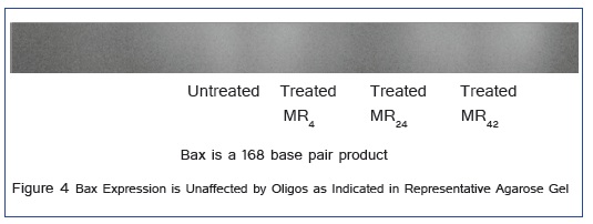
When background intensity was subtracted, the relative intensity of the bands corresponding to bax representing cells treated with MR4, MR24 and MR42 compared to controls were -5.74 ± 16.9, 5.54 ±19.2, and -15.34 ± 32.9. These results were pooled from both duplicate PCR runs and gels and indicated that no significant differences in bax expression were found, compared to that seen with BCL-2.
Caspase-3 expression
Comparable amounts of extracted RNA from LNCaP cells treated with either mono- or bispecific oligos directed against BCL-2 (and EGFR in the bispecifics) was evaluated by RT-PCR using primers directed against caspase-3. A representative band for caspase-3 is presented in Figure 5 and appears immediately below the marker representing 300 base pairs.

When background intensity was subtracted, the relative intensity of the bands corresponding to caspase-3 representing cells treated with MR4, MR24 and MR42 compared to controls were -35.8 ± 12.5 (P = 0.0002), -40.3 ± 16.6 P = 0.0006) and -43.5 ± 26.3 (P = 0.006). These results were pooled from both duplicate PCR runs and gels and indicated that similar significant suppression of caspase-3 activity was demonstrated with each oligo (and type) evaluated. This result would suggest that when inhibiting BCL-2, caspase-3 activity should be either maintained or replaced.
Clusterin expression
Comparable amounts of extracted RNA from LNCaP cells treated with either mono- or bispecific oligos directed against BCL-2 (and EGFR in the bispecifics) was then evaluated by RT-PCR using primers directed against clusterin. A representative band for clusterin is presented in Figure 6 and appears where expected between the markers representing 200 and 300 base pairs, as a 221 base pair product.

When background intensity was subtracted, the relative intensity of the bands corresponding to clusterin representing cells treated with MR4, MR24 and MR42 compared to controls were 8.3% ± 14.5, 9.0% ±17.3, and -14.1% ± 22.6 (mean ± SD). These results were pooled from both duplicate PCR runs and gels, indicating (like bax) there are no significant differences in clusterin expression, compared to that seen with caspase-3.
AKT-1 expression
Comparable amounts of extracted RNA from LNCaP cells treated with either mono- or bispecific oligos directed against BCL-2 (and EGFR in the bispecifics) was then evaluated by RT-PCR using primers directed against AKT-1. A representative band is presented in Figure 7 and appears as a 189 base pair product.

When background intensity was subtracted, the relative intensity of the bands corresponding to AKT-1 representing cells treated with MR4, MR24 and MR42 compared to controls were increased 256.7% ± 105.5 (P = 0.0006), 189.4% ±73.6 (P = 0.0004), and 182.6% ± 90.8 (P = 0.002) (mean ± SD). These results indicate that the expression of the apoptotic inhibitory protein AKT-1 is enhanced, identifying another compensatory response to BCL-2 suppression.
MED-12 expression
Comparable amounts of extracted RNA from LNCaP cells treated with either mono- or bispecific oligos directed against BCL-2 (and EGFR in the bispecifics) was then evaluated by RT-PCR using primers directed against MED-12. A representative band is presented in Figure 8 and appears as a 183 base pair product.

When background intensity was subtracted, the relative intensity of the bands corresponding to MED-12 representing cells treated with MR4, MR24 and MR42 compared to controls were increased 138.1% ± 120.1 (P = 0.018239), 181.3% ±165.4 (P = 0.022919), and 61.4% ± 126.5 (NS) (mean ± SD). These results suggest that the expression of the transcription factor protein MED-12 is enhanced by at least some mono- and bispecific oligos directed against BCL-2 and identify yet another compensatory response.
PD-L1 expression
Comparable amounts of extracted RNA from LNCaP cells treated with either mono- or bispecific oligos directed against BCL-2 (and EGFR in the bispecifics) was then evaluated by RT-PCR using primers directed against PD-L1. A representative band is presented in Figure 9.

When background intensity was subtracted, the relative intensity of the bands corresponding to PD-L1 representing cells treated with MR4, MR24 and MR42 compared to controls were increased 33.3% ± 20.1 (P = 0.006079), 51.0% ±36.7 (P = 0.014394), and 28.5% ± 19.3 (P = 0.010793) (mean ± SD). These results indicate that the expression of the immune blockade marker PD-L1 is significantly enhanced (although not to a great extent), suggesting once again another compensatory response to BCL-2 suppression may occur.
Discussion
Gene therapy for cancer presents a far more complex challenge than treating single gene inherited deficiencies because for it to work numerous pathways (and many of their regulatory proteins) must be simultaneously regulated (suppressed or replaced). Potentially hundreds or even thousands of genes may be involved or having altered expression patterns. The challenge is to identify those gene products which are critical to either continuing the process of malignant transformation or those which maintain normal differentiated function.
For gene products which are overexpressed, methods to suppress either their translation or activities have been developed, including the use of antisense oligos. For the treatment of prostate cancer, some (produced by Oncogenex Pharmaceuticals) have reached clinical trials (OGX-011), while others continue their preclinical development (OGX-225). Often administered in combination with traditional chemotherapy, these oligos target proteins which include BCL-2, clusterin (OGX-011 in Phase II testing), heat shock protein 27 (OGX-427) and insulin growth factor binding proteins (OGX-225).The most promising approach involves efforts to restore tumor apoptosis by eliminating proteins associated with this aspect of tumor resistance.
For those proteins diminished or lacking in expression gene replacement, promotion or amplification would be necessary. Although not as far advanced as gene suppressive therapy, suppressor gene PTEN has been replaced through adenoviral transfection in a non-small cell lung cancer model, where it restored effective radiation treatment through diminished capacity for DNA repair [12]. In a prostate cancer model PTEN activity is associated with suppression of BCL-2 and increased chemosensitivity [13]. In addition, antisense treatment directed against BCL-2 increases radiosensitivity in both prostate [14] and nasopharyngeal [15] cancers.
Tumor cells are heterogenous and those which evade growth regulation or apoptosis are positively selected. In addition, variants, as their DNA becomes increasingly unstable, tend to accumulate additional adaptations (mutations) which further contribute to resistance and dissemination.
Treatment protocols administered to correct one genetic alteration can, through selective pressure, initiate compensatory changes which diminish the effectiveness of the original, and in many tumors, some of the early mutational events lead to evasion of apoptosis. This ?programmed? process clears the body of altered (transformed) or damaged cells. It is highly regulated and involves many proteins being synthesized, recognized (via receptors) or otherwise interacting with each other. Selection of cells which resist apoptosis is no different than the process by which hormone sensitive prostate tumor cells, in the absence of androgen, are selected (and establish themselves) as insensitive variants. Therefore, for suppressive gene therapy to work, it?s important to identify compensatory effects.
Previous work identified increased androgen sensitivity as one mechanism compensating for BCL-2 suppression. In this study we evaluated the effect of oligo mediated growth suppression on various inhibitors (BCL-2, clusterin, AKT-1) and promoters (bax, caspase-3) of apoptosis and found that within this pathway at least two proteins (caspase-3 and AKT-1) compensate for BCL-2 suppression.
We conclude that this study identifies a new form of tumor resistance which results in a compensatory evasion of apoptosis, in addition to alterations in gene transcription and immune activation. Together with the previously identified increased androgen sensitivity which produces a gene expression pattern often seen in later stage tumors, suppressive BCL-2 directed therapy could result not only in restored apoptosis, but increased androgen stimulation and tumor aggressiveness.
For gene therapy to be successful, additional effects on untargeted genes must be identified. This is particularly important when translational inhibitors are administered and directed against BCL-2 (particularly for the treatment of prostate cancer). Should additional proteins (inhibitors of apoptosis) be indicated for suppression, bispecific oligos or even proposed multifunctional and branched derivatives could then be employed [16]. Additional studies are underway to identify altered expressions in other proteins associated with apoptosis.
Conflict of Interest Statement
None; The authors have no conflict of interest or financial reward in the results of this study.
Acknowledgments
The Cellular Biology laboratory at the Hektoen Institute is supported, in part, by the Blum Kovler Foundation, the Cancer Federation, and Safeway/Dominicks Campaign for Breast Cancer Awareness, Lawn Manor Beth Jacob Hebrew Congregation, the Max Goldenberg Foundation, the Sternfeld Family Foundation, and the Herbert C. Wenske Foundation.